Novel PET tracer detects synaptic changes in spinal cord and brain after spinal cord injury
. DOI: 10.2967/jnumed.124.269291")
A new PET tracer can provide insights into how spinal cord injuries affect not only the spinal cord, but also the brain, according to new research published in The Journal of Nuclear Medicine. By identifying synapse loss, the PET approach provides molecularly unique and complementary information to other structural imaging methods, offering a promising objective metric to evaluate novel therapeutics for spinal cord injuries.
According to the National Spinal Cord Injury Statistical Center, the annual incidence of traumatic spinal cord injury is about 54 cases per 1 million people, and approximately 308,600 people in the United States live with a spinal cord injury. Clinical outcomes vary based on injury severity and location, potentially leading to partial or complete loss of sensory or motor function below the injury level. Current clinical spinal cord injury diagnosis relies on anatomic techniques such as X-ray and CT, which assess spinal integrity but provide limited physiologic and pathologic information.
“There is an urgent need for a quantitative and noninvasive imaging method for neural network changes after spinal cord injury,” said Jason Cai, Ph.D., associate professor of radiology and biomedical imaging and of pharmacology at Yale School of Medicine in New Haven, Connecticut.
“By offering a noninvasive quantitative method to visualize and quantify synapse loss in the whole central nervous system, SV2A PET could become an essential tool for evaluating and monitoring the progression of spinal cord injury or predict recovery.”
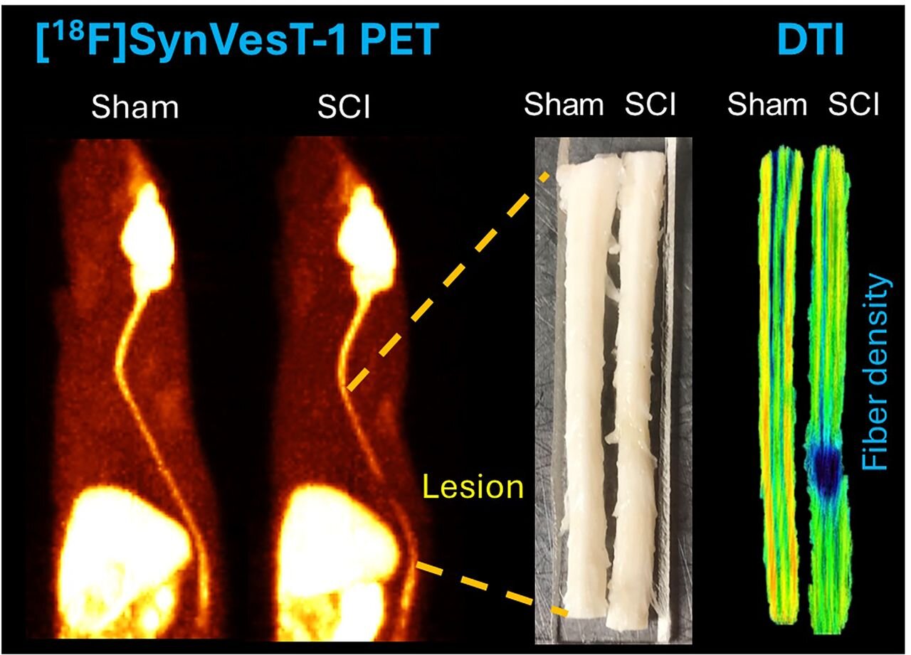
Researchers used the newly developed 18F-labeled SV2A radiotracer, [18F]SynVesT-1, to assess changes in synaptic density in a rat model of T7 contusion. Nine rats with T7 spinal cord injuries and seven sham controls were imaged with [18F]SynVesT-1 PET on day 1 and on days 9 through 11 after injury. Imaging findings of the injury site and of the brain were compared with ex vivo diffusion tensor imaging (DTI) and molecular biologic analyses.
[18F]SynVesT-1 PET effectively identified synapse loss in the contusion SCI rat model. Uptake at the spinal cord injury epicenter was found to be reduced by 58% and 52% on day one and days nine through 11 after injury, respectively, compared with the sham control rats. The uptake of 18F-SynVesT-1 in the amygdala and cerebellum was also lower in spinal cord injury rats, and ex vivo DTI analysis revealed fiber damage in the internal capsule and somatosensory cortex.
“Our work has potential to revolutionize the way spinal cord injury is diagnosed and monitored in the clinic,” noted Cai. “SV2A PET could be used to evaluate the effects of new treatments objectively and quantitatively, supporting more precise and personalized therapeutic strategies for patients with spinal cord injuries.”
More information:
Baosheng Chen et al, [18F]SynVesT-1 PET Detects SV2A Changes in the Spinal Cord and Brain of Rats with Spinal Cord Injury, Journal of Nuclear Medicine (2025). DOI: 10.2967/jnumed.124.269291
Provided by
Society of Nuclear Medicine and Molecular Imaging
Citation:
Novel PET tracer detects synaptic changes in spinal cord and brain after spinal cord injury (2025, September 12)
retrieved 12 September 2025
from https://medicalxpress.com/news/2025-09-pet-tracer-synaptic-spinal-cord.html
This document is subject to copyright. Apart from any fair dealing for the purpose of private study or research, no
part may be reproduced without the written permission. The content is provided for information purposes only.